Protein Data Bank, PDB
This week our class runs as scheduled and now we learn about PDB, some sort of 3-D micro-molecular structure. Using RasMol version 2.6 for viewing gives quite restricted command as i dont get any command line to make the creature moves. We are asked to get two structure of trypsin from the RSCB web site.if im not mistaken, and well..the internet seem make all things slow and slower...
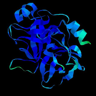
Then, here's the image of the structure using RasMol, crystal structure of anionic trypsin isoform 3 from chum salmon (blue one) using the ribbon display with temperature colouring. The information of the structure can be get at here.

Then, here's the image of the structure using RasMol, crystal structure of anionic trypsin isoform 3 from chum salmon (blue one) using the ribbon display with temperature colouring. The information of the structure can be get at here.

The second structure (right) is Anionic trypsin in complex with bovine pancreatic trypsin inhibitor (BPTI) which is found in the Rattus norvegicus or commonly rat. The information of it can be found here.
This structure is viewed in the ball and stick display and shapely coloured.
This structure is viewed in the ball and stick display and shapely coloured.

0 comments:
Post a Comment